Refractive Surgery & Corneal Modification Defs
AberrometryA method of capturing the wavefront of an ocular system. Typically a light beam is projected into the eye and the aberrometer captures the existing rays as they are reflected off of the retina. The wavefront profile of the eye is then displayed in a two-dimensional or three-dimensional map. Usually five measurements are taken and the average of the three closest readings produces the final wavefront measurement. Wavefront maps are displayed in terms of Zernike polynomials and measured in microns.
Ablate
In surgery, to remove.
Astigmatic Keratotomy (AK)
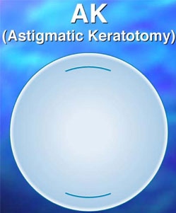
Incisional surgical procedure used to correct corneal astigmatism. Arcuate incisions are placed in the corneal midperipheral zone of the steep meridian at approximately 90% depth.
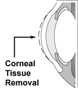
Automated Lamellar Keratoplasty (ALK)
A refractive surgical procedure in which the surgeon creates a flap of the uppermost layer of the cornea using a microkeratome. A second pass of the microkeratome is made in order to remove a wedge of tissue.
Broad Beam Laser
Excimer laser where the beam size used to ablate the cornea is from approximately 6.0 to 8.0 millimeters.
Conductive Keratoplasty (CK)
CK is a non-ablative, collagen-shrinking procedure for the treatment of mild and moderate hyperopia. Radiofrequency energy is delivered through a fine tip inserted into the corneal stroma. The collagen lamellae in the area surrounding the tip shrink and tighten, increasing the radius of curvature of the cornea. The spots are placed in the circumference of the mid- and peripheral cornea. Based on the amount of refractive change targeted, the number and location of treatment spots may be determined, with larger treatments requiring more spots and rings.
Conventional Refractive Surgery
Method of optical correction by Excimer laser photoablation, which changes the shape of the cornea to change the refraction error. The treatments are symmetrical and correct lower order or spherocylindrical aberrations, which include myopia, hyperopia, and astigmatism.
Current treatment parameters: Myopia 0.50 diopters to 12.00 diopters
Hyperopia 0.50 diopters to 6.00 diopters
Astigmatism 0.50 diopters to 6.00 diopters
Customized Refractive Surgery
Wavefront-driven excimer laser photoablation, which changes the shape of the cornea to change refractive error. In addition to treating lower-order aberrations (sphere and cylinder), custom refractive surgery treats higher-order aberrations up to the 6th order. Wavefront-guided ablations create treatment profiles based on wavefront maps and the treatment may be asymmetrical and is customized for the individual patient. The treatment parameters vary according to the system utilized. The first customized laser platform was FDA approved in the fall of 2002.
Ectasia
Progressive corneal thinning and associated protrusion.
Epi-LASIK
Refractive surgical procedure.
This new procedure is believed to avoid risks associated with laser-assisted in situ keratomileusis (LASIK) and offers improved postoperative recovery compared with Photorefractive Keratectomy (PRK). Rather than creating a flap with a microkeratome as in LASIK or removing the epithelium as in PRK, Epi-LASIK separates an epithelial sheet using the Centurion SES EpiEdge epikeratome (CIBA Vision).
Epithelial Ingrowth
A LASIK complication wherein epithelial cells proliferate underneath the corneal flap.
Excimer Laser
Type of laser used in all laser refractive surgical procedures in order to reduce refractive error. The laser utilizes a 193 nm argon-fluoride beam to reshape the anterior corneal stroma by breaking collagen bonds and expelling or ablating corneal tissue with each laser pulse. It is termed a "cold" laser because the collagen bonds are broken without damaging adjacent cells. The pulses are fired centrally to flatten the cornea to an oblate shape in myopia and fired peripherally to steepen the cornea to a prolate shape in hyperopia.
Femtosecond Laser
High-frequency laser now used to make flaps in the LASIK procedure. The high-frequency energy can be focused through the anterior cornea to a specified depth. A sweeping back and forth (raster) pattern creates a horizontal, then vertical cleavage plane to create the flap.
Intrastromal Corneal Ring Segments (ICRS)
A reversible procedure used in the treatment of low amounts of myopia (-1.00 to -3.00 D) by placing rings of polymethylmethacrylate (PMMA) in the midperipheral corneal stroma to flatten the radius of curvature. Intrastromal corneal ring segments are currently being investigated to reduce myopia in patients with keratoconus and corneal ectasia.
Keratomileusis
The sculpting of the cornea, formerly done with a lathe and blade, now done with an excimer laser.
Keratoplasty
The replacement of the cornea. Keratoplasty can be lamellar (replacement of the superficial layers) or penetrating (transplantation or replacement of the full thickness of the cornea).
Keratotomy
A surgical incision of the cornea.
LASER
An acronym for light amplification by stimulated emission of radiation.
Laser-Assisted In Situ Keratomileusis (LASIK)
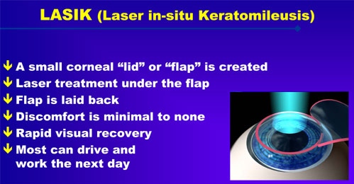
Approved by the FDA for the correction of myopia, hyperopia and astigmatism. LASIK is a combination of two refractive technologies: Use of a microkeratome, to create a thin flap of tissue (approximately 130 to 180 microns thick) followed by excimer laser ablation to reshape the stromal tissue beneath the flap.
Laser Epithelial Keratomileusis (LASEK)
A hybrid of photorefractive keratectomy (PRK) and laser-assisted in situ keratomileusis (LASIK), the goal of LASEK is the preservation of the corneal epithelium. Rather than creating a flap with a microkeratome (as in LASIK) or scraping and removing the patient's epithelium (as in PRK), LASEK treats the epithelium with alcohol to loosen and separate it from the stroma and it is then rolled back. The underlying stroma is ablated with an excimer laser and the epithelial cells are rolled back out, repositioned, and smoothed. The potential advantages of LASEK are to reduce postoperative haze, speed visual recovery, and decrease postoperative pain over traditional PRK.
Laser Thermal Keratoplasty (LTK)
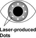
Approved by the FDA in 2000 for the temporary correction of hyperopia ranging from +0.75 to +2.50 D with less than 1.0 D of astigmatism. LTK uses a Holmium:YAG laser comprised of a slit-lamp delivery system that creates spots around the circumference of the peripheral cornea. The treatment consists of either one or two rings of spots set at an optical zone of 6.0 and/or 7.0 mm.
Micron
One thousandth of a millimeter. One millionth of a meter.
Microkeratome
Instrument used for the creation of a lamellar flap (130 to 180 mm) during laser-assisted in situ keratomileusis (LASIK). A microkeratome is comprised of a suction ring that adheres to the globe, providing stability for the cutting blade that rolls along a tongue and groove track, creating the flap.
Nomogram
The surgical adjustment to a laser's computer calculation to further refine results.
Pachymetry
Measurement of corneal thickness. Methods of measurement are based on wave reflection of optical light or ultrasonic energy through the corneal tissue.
Photorefractive Keratectomy (PRK)
A procedure involving the removal of the epithelium by gentle scraping away of the corneal epithelium and use of a computer-controlled excimer laser to reshape the stroma.
Phototherapeutic Keratotomy (PTK)
PTK involves ablative photodecomposition of the epithelium by ablating microscopically thin layers and etching away surface irregularities. Candidates for PTK are patients with significant visual compromise due to corneal scars and opacities (from trauma or inactive infections), dystrophies (Reis-Buckler's, lattice, anterior basement membrane dystrophy [ABMD]), irregular corneal surface associated with filamentary keratitis and Salzmann's nodular degeneration, recurrent corneal erosions (RCE) (unresponsive to lubricants, debridement, or stromal puncture), band keratopathy, scars resulting from previous pterygium excision, Thygeson's superficial keratitis, and irregular astigmatism.
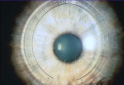
Radial Keratotomy (RK)
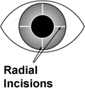
Refractive surgical procedure popular in the 1980s, RK treated low amounts of myopia ranging from -1.00 to -4.00 D. In order to flatten the cornea, radial incisions (like the spokes of a wheel) were created using a diamond-blade micrometer knife, leaving a central unaltered optical zone of approximately 3.0 mm.
Refractive Surgery
A surgical method of vision correction by changing the refractive properties of the eye.
Scleral Expansion Bands (SEB)
Procedure potential is for the reversal of presbyopia. Increasing lens zonular tension by implanting small polymethylmethacrylate (PMMA) bands in the sclera over the ciliary body will allow accommodation to occur.
Wavefronts
Wavefront capture by an aberrometer is the measurement of lower- and higher-order aberrations. As a wavefront passes through multiple refractive surfaces, ocular aberrations are induced that prevent individual light beams from focusing at the same point on the macula. Ocular aberrations increase as a function of pupil size and become visually significant with pupil dilation. In a perfect optical system the wavefront would be flat. Optical aberrations are as unique as a person's fingerprint, with each eye producing its own unique wavefront . Once a patient's wavefront is captured, it is incorporated into the refractive surgical procedure for a customized treatment.
Content supplied by the AOA Contact Lens and Cornea Section

Special thanks to Ann Laurenzi, O.D., Contact Lens and Cornea Section's Refractive Surgery Technology Committee Chair for her contributions to this content.